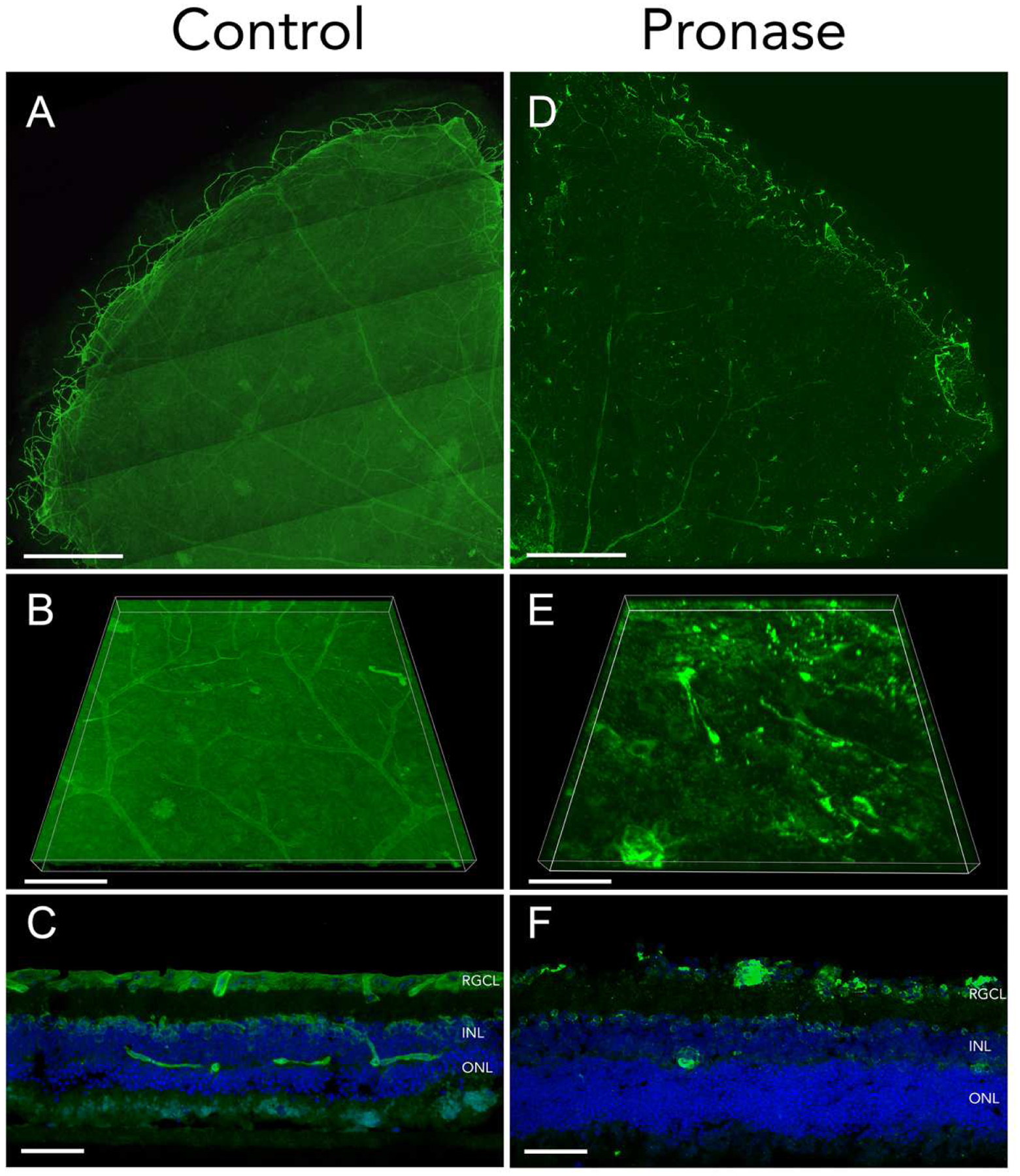
Figure 2. Effects of Pronase-E digestion on ILM laminin.

Adult organotypic mouse retinal explants treated with BSS (A-C) or Pronase-E at a concentration of 0.6 U/mL (D-F) were cultured for 7 days. Fixed tissue stained with laminin (green) shows ILM organization. Tiled confocal microscopy of flat mount retina quadrants are shown (A, D). Three-dimensional reconstruction of confocal microscopy z-stacks detail ILM surfaces in control (B) and Pronase (E) treated retinas. Retinal explant cryosections exhibit intact (C) and linear interruptions (F) in ILM, with preserved retinal layers indicated by DAPI counterstain of retinal cell nuclei in blue. Scale bars: 500μm (A, D); 100μm (B, E); 50μm (C, F).
