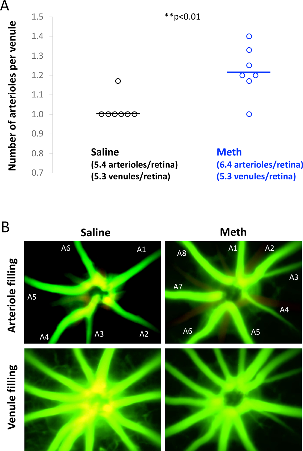
Figure 2.

A. Number of retinal arterioles per venule in control (saline-) and METH-injected mice at Day 26. N=7 per group. **p<0.01. B. Example of arteriolar and venular filling of fluorescent dye to determine the number of primary arterioles (filling first; numbered A1-A7) and venules (filling subsequently).
