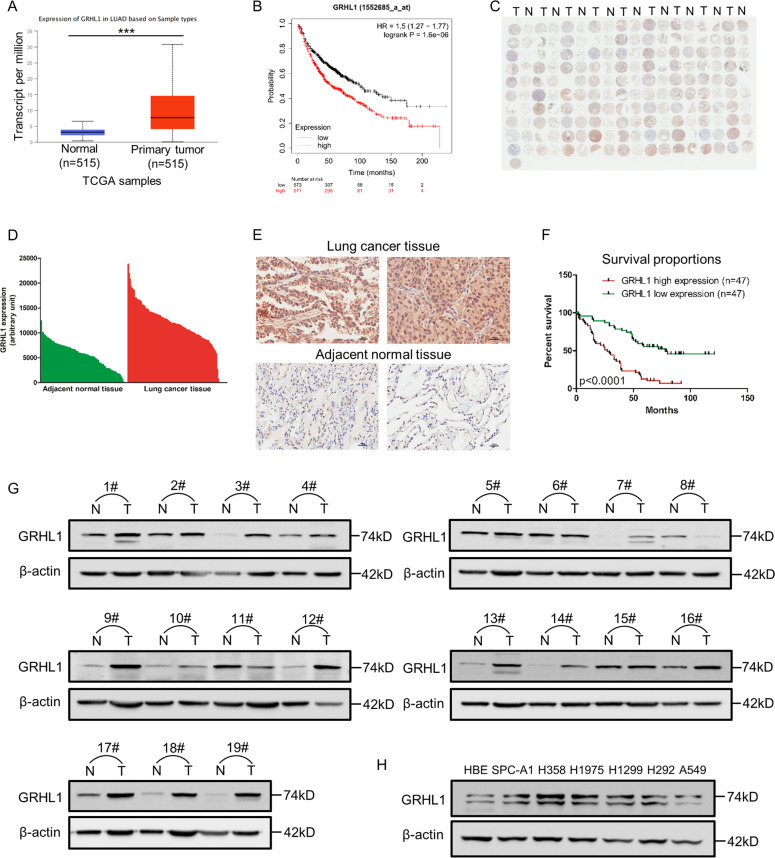
Fig. 1. GRHL1 is upregulated in NSCLC and closely related to the poor survival of NSCLC patients.
A The mRNA levels of NSCLC samples (Normal and Primary tumor) were analyzed from TCGA using the following link: http://ualcan.path.uab.edu/cgi-bin/ualcan-res.pl. ***: P < 0.0001. B The overall survival curves of NSCLC patients were analyzed using Kaplan–Meier Plotter (https://kmplot.com/analysis/). C Immunohistochemical staining of a representative lung adenocarcinoma tissue microarray with an anti-GRHL1 antibody. T, tumor tissue; N, adjacent normal tissue. D Quantification of the immunohistochemical (IHC) staining shown in Fig. 1C. E Microscopic evaluation of IHC staining of two representative tumor tissues and adjacent normal tissues shown in Fig. 1C with an anti-GRHL1 antibody (brown) and hematoxylin counterstain (blue). Scale bars: 100 μm. F Kaplan–Meier survival curve of 94 NSCLC patients. Patients were divided into two groups according to the average staining density of GRHL1 in cancer tissues of the tissue array (high expression: n = 47, low expression: n = 47, Log-rank (Mantel-Cox) test was used for the statistical analysis). G The protein expression levels of GRHL1 were determined by western blot using the paired, tumor-adjacent noncancerous lung tissues (normal, N) and human NSCLC tissues (tumor, T) from 19 NSCLC patients (1#−19#). H The expression levels of GRHL1 in NSCLC cell lines (SPC-A1, H358, H1975, H1299, H292, A549) and HBE cells were examined by western blot using an anti-GRHL1 antibody.

