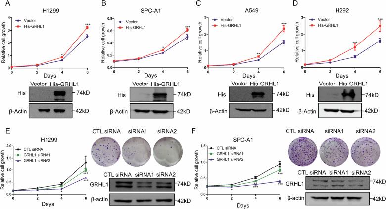
Fig. 2. GRHL1 promotes the proliferation of NSCLC cells.
A–D H1299 (A) cells, SPC-A1 (B) cells, A549 (C) cells, and H292 (D) cells were transfected with pcDNA3.1 plasmid or pcDNA3.1-His-GRHL1 (His-GRHL1) plasmid. At the indicated times, cells were fixed in 3.7% formaldehyde and stained with 0.1% crystal violet. Data represent the average of three independent experiments (mean ± SD). *P < 0.05, ***P < 0.001. Western blot was used to examine the expression of the indicated proteins. E, F H1299 (E) and SPC-A1 (F) cells were transfected with control (CTL) siRNAs or GRHL1 siRNAs. At the indicated times, cells were fixed in 3.7% formaldehyde and stained with 0.1% crystal violet. Data represent the average of three independent experiments (mean ± SD). *P < 0.05, **P < 0.01, ***P < 0.001. For colony formation assay, H1299 and SPC-A1 cells were transfected with control siRNA or siRNAs targeting GRHL1. Eight days later, colony formation was detected by crystal violet staining and photographed. Protein expression was assessed by western blot using indicated antibodies.

