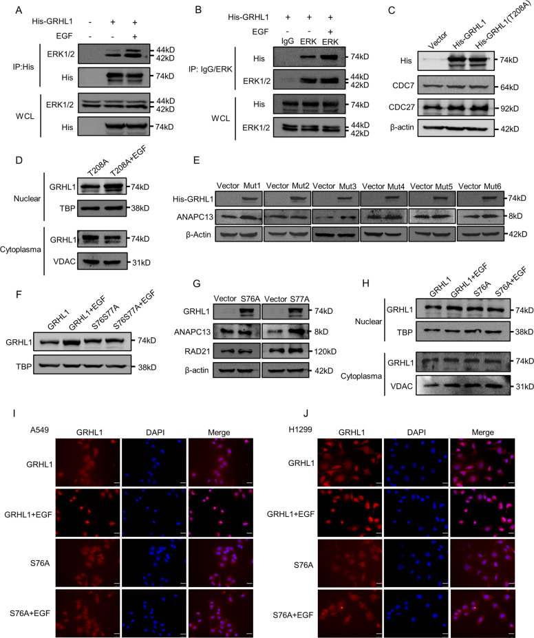
Fig. 6. ERK is responsible for the phosphorylation of GRHL1 at Ser76.
A 293 T cells were transfected with control plasmid and His-GRHL1 plasmid. After serum-free treatment for 12 h and 12 h of stimulation with EGF (100 ng/ml), cells were lysed for immunoprecipitation using an anti-His antibody and blotted with indicated antibodies. B 293 T cells were transfected with His-GRHL1. After serum-free treatment for 12 h and 12 h of stimulation with EGF (100 ng/ml), cells were lysed for immunoprecipitation using anti-ERK1/2 antibody and control rabbit IgG and blotted with indicated antibodies. C A549 cells were transiently transfected with control plasmid, His-GRHL1 plasmid and His-GRHL1(T208A) plasmid respectively. Forty-eight hours later, the cells were lysed. Protein expression was assessed by western blotting using the indicated antibodies. D A549 cells were transfected with His-GRHL1(T208A) plasmid, after serum-free treatment for 12 h and 24 h of stimulation with EGF (100 ng/ml), the nuclear and cytoplasmic fractions were separated and the location of GRHL1 was detected by western blot. E A549 cells were transiently transfected with control plasmid and six mutants (mut1: S25S28S35A, mut2: S76S77A, mut3: S95A, mut4: S442A, mut5: S493S501A, mut6: S539A) plasmids respectively. Forty-eight hours later, the cells were lysed. Protein expression was assessed by western blotting using the indicated antibodies. F A549 cells were transfected with His-GRHL1 plasmid and His-GRHL1(S76S77A) plasmid, after serum-free treatment for 12 h and 24 h of stimulation with EGF (100 ng/ml), the nuclear and cytoplasmic fractions were separated and the GRHL1 in nuclear was detected by western blot. G A549 cells were transiently transfected with control plasmid, His-GRHL1(S76A), and His-GRHL1(S77A) plasmids respectively. Forty-eight hours later, the cells were lysed. Protein expression was assessed by western blotting using the indicated antibodies. H A549 cells were transfected with His-GRHL1 plasmid and His-GRHL1(S76A) plasmid, after serum-free treatment for 12 h and 24 h of stimulation with EGF (100 ng/ml), the nuclear and cytoplasmic fractions were separated and the location of GRHL1 was detected by western blot. I–J A549 and H1299 cells were transfected with His-GRHL1 plasmid and His-GRHL1(S76A) plasmid. After serum-free treatment for 12 h, EGF (100 ng/ml) was added. After 12 h. The location of GRHL1 was detected by immunofluorescence staining. Cells were stained with anti-GRHL1 (left panel, red) and DAPI (middle panel, blue). The merged images are shown in the right panel. Scale bar = 25 µm, magnification: ×400.

