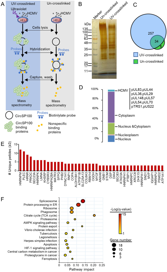
Fig. 5.
Analysis of circSP100 binding proteins captured by RAP. A The strategy of RAP mass spectrometry; B Proteins purified with RAP from UV-crosslinked or un-crosslinked lysis extracted from HCMV infected HELF cells; C Venn diagrams of proteins purified in RAP; D Subcellular location of the proteins that interacted directly with circSP100; E The circSP100 binding proteins. Only the proteins of unique peptides ≥ 3 were indicated; F KEGG enrichment of the proteins that interacted with circSP100.

