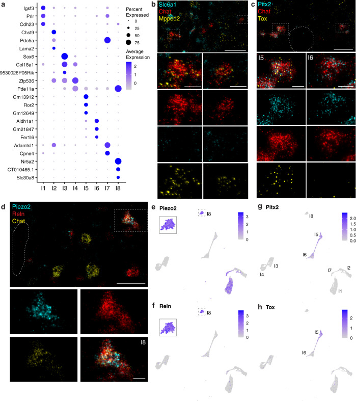
Fig. 5. Spinal cholinergic interneurons separate into 8 transcriptionally distinct subtypes.
a Top markers of the 8 cholinergic interneuron clusters. b ISH showing expression of two top markers for cholinergic interneurons (Fig. 2c), Slc6a1 (cyan) and Mpped2 (yellow) in Chat+ neurons (red) in the intermediate zone. Boxes highlight a neuron expressing Chat and Mpped2, but lacking Slc6a1 (left), and a neuron expressing Chat, Mpped2, and Slc6a1 (right). c ISH showing expression of Pitx2 and Tox near the central canal (outlined) of the lumbar spinal cord. Co-expression of Pitx2 and Tox in Chat+ cells exemplifies cluster I5 (left) whereas Pitx2 and Chat without Tox identify a cluster I6 cell (right). d High-magnification images showing co-expression of Piezo2, Reln, and Chat in an I8 neuron found near the central canal (outlined) of the thoracic spinal cord. e, f Piezo2 and Reln co-expression are diagnostic of a rare population of interneurons, cluster I8. g Pitx2, a known marker for partition cells, is expressed only in clusters I5 and I6. h Tox is expressed only in interneuron cluster I5 and thus differentiates between the two Pitx2-expressing clusters. Low magnification scale bars, 50 µm. High magnification scale bars, 10 µm.

