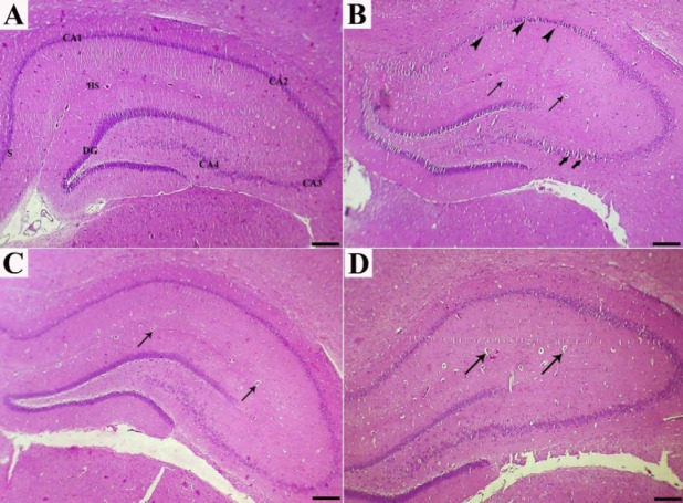
Figure 1.
Sections of the hippocampus in experimental rats, as stained by Hematoxylin and Eosin: (a) In normal rats, the hippocampus consists of CA1, CA2, CA3, and CA4 areas; CA1 is continued as subiculum (S). hippocampus sulcus (HS) and dentate gyrus (DG) are narrow. (b) cholesterol-rich diet rats showed prominent reduction in pyramidal cells accompanied by presence of a few shrunken degenerated cells (arrowheads) and vascularization (thin arrows). Slight spongiosis (vacuolations) was also observed in the CA4 area (thick arrows). (c and d) High-cholesterol diet-fed rats treated with dill tablets and aqueous extract of basil showed a better organization in the hippocampus layers and decreased number of capillaries (arrows) with moderate gliosis. Bar=500 µm

