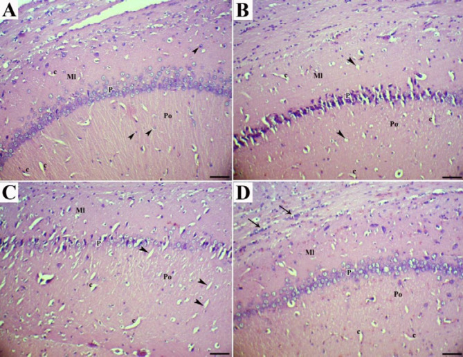
Figure 2.
CA1 area of the hippocampus (H&E; Bar=60 µm). (a) normal rats showed molecular (Ml), pyramidal (P), and polymorphic (Po) layers. As shown by arrowheads, the glial cells and capillaries (c) were found irregularly distributed in molecular and polymorphic layers. (b) High cholesterol diet-fed rats showed marked shrinkage in the size of large pyramidal cells, necrotic pyramidal cells with eccentric nuclei dispersed small vacuolations (arrowheads), and vascularization (c). (c) High cholesterol diet-fed rats treated with dill tablets showed less degenerative and necrotic pyramidal cells, moderate gliosis in the molecular layer, and presence of capillaries and small vacuolations in the molecular layer (arrowheads). (d) High-cholesterol diet-fed rats treated with aqueous extract of basil showed pyramidal cells with normal size and shape but clumping of nuclei and neuronal processes and massive gliosis in the molecular layer (arrows)

