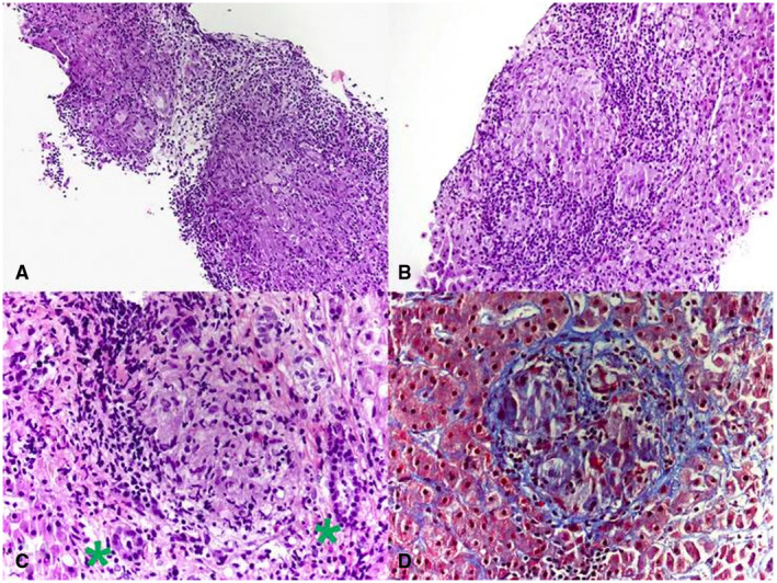
FIG 2.

Histology of sarcoid granuloma. Photomicrograph from liver biopsy showing multiple coalescing nonnecrotizing granulomas (hematoxylin and eosin, original magnification 10×). (B) Lobular nonnecrotizing granuloma with tightly packed epithelioid cells surrounded by lymphocytes (hematoxylin and eosin, original magnification 20×). (C) Portal nonnecrotizing granuloma adjacent to uninvolved bile ducts (green asterisks) (hematoxylin and eosin, original magnification 40×). (D) Mild perigranulomatous fibrosis (Trichrome stain, original magnification 40×). Reproduced from Frontiers in Medicine. 11 Copyright 2019, Creative Commons Attribution 4.0.
