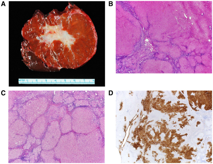
FIG 2.

FNH. (A) Gross image of a typical FNH demonstrating a central scar. (B) The lesion (lower left) is distinct from the background liver (upper right) but is not encapsulated. (C) The lesion is composed of nodules of benign hepatocytes separated by fibrous septa that contain inflammation and ductular proliferation. (D) A GS immunostain shows a characteristic map‐like staining pattern, with interconnecting broad bands of strongly staining hepatocytes, but with no staining of hepatocytes adjacent to the fibrous bands. A small portion of normal liver, with normal zone 3 perivenular staining of hepatocytes, is seen on the left side of the image.
