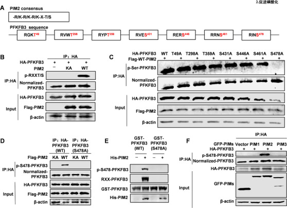
FIGURE 2.

PIM2 phosphorylates PFKFB3 at Ser478. (A) Schematic diagram of sites in PFKFB3 that may be modified by PIM2. (B) HEK293T cells were overexpressed the indicated HA‐PFKFB3 and Flag‐PIM2 (WT or KA) proteins. Immunoprecipitation with an anti‐HA antibody was performed. (C) HEK293T cells were overexpressed the indicated HA‐PFKFB3 (WT or mutant) and Flag‐PIM2 (WT). Immunoprecipitation with an anti‐HA antibody was performed. (D) HEK293T cells were cotransfected with HA‐PFKFB3 (WT or S478A) and Flag‐PIM2 (WT or KA) proteins. Immunoprecipitation with an anti‐HA antibody was performed, followed by Western blot with indicated antibodies. (E) Purified GST‐PFKFB3 was mixed with the indicated bacterially purified His‐PIM2 proteins. An in vitro kinase assay was performed. (F) HEK293T cells were overexpressed the indicated HA‐PFKFB3 and GFP‐PIMs (PIM1, PIM2, PIM3). Immunoprecipitation with an anti‐HA antibody was performed. All experiments were repeated at least 3 times
