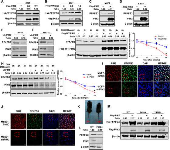
FIGURE 3.

PIM2 regulates PFKFB3 protein stability. (A) HEK293T cells were cotransfected with HA‐PFKFB3 and Flag‐PIM2 (WT or KA) plasmid. Western blot analysis tested whole‐cell lysate after 72‐h transfection. (B) HEK293T cells were cotransfected with HA‐PFKFB3 and Flag‐PIM2 (0, 0.5, 1.0 μg) plasmid. Western blot analysis tested whole‐cell lysate after 72‐h transfection. (C and D) MCF7 or MB231 cells were cotransfected with Flag‐PIM2 plasmid. Whole‐cell lysate after 72‐h transfection. (E and F) MCF7 or MB231 cells were knocked down PIM2 with shRNA. Total cell lysates were prepared. (G) MCF7 cells were transfected with HA‐PFKFB3 and Flag‐PIM2 plasmids, and then treated with CHX for indicated time. Total cell lysates were prepared. (H) MCF‐7 cells with stable knockdown PIM2 proteins were treated with CHX for indicated time. Total cell lysates were prepared. (I and J) MCF7 or MB231 cells were knocked down PIM2 with shRNA. Confocal immunofluorescence microscopy was performed to observe the expression of PIM2 and PFKFB3. Red box mark was partially enlarged in the Figures S1B and S1C. (K) PIM2 gene knockout mice constructed and screened. (L) Detection of PIM2 and PFKFB3 expression in PIM2 knockout mice by WB. (M) MCF‐7 cells were overexpressed the indicated both Flag‐PIM2 and HA‐PFKFB3 (WT, S478A, or S478D) proteins. Total cell lysates were prepared. All experiments were repeated at least 3 times
