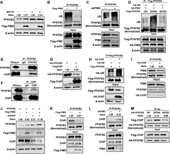
FIGURE 4.

PIM2 promotes PFKFB3 protein stability via the CHIP‐mediated ubiquitin proteasome pathway. (A) MCF7 cells were cotransfected with Flag‐PIM2, and HA‐PFKFB3 plasmids were treated with or without MG132 for 8 h. Total cell lysates were prepared. (B and C) MCF7 cells were cotransfected with Flag‐PIM2 or sh‐PIM2. Immunoprecipitation with an anti‐PFKFB3 antibody was performed. (D) MCF7 cells were co‐overexpressed the indicated HA‐PIM2 and Flag‐PFKFB3 (WT, S478A or S478D) proteins. Immunoprecipitation with an anti‐Flag antibody was performed. (E and F) Immunoprecipitation (IP) and immunoblotting (IB) analyses were performed with indicated antibodies in MCF7. (G) MCF7 cells were cotransfected with HA‐PFKFB3 and Flag‐CHIP plasmid. Western blot analysis tested whole‐cell lysate after 72‐h transfection. (H) MCF7 cells were co‐overexpressed the indicated HA‐CHIP and Flag‐PFKFB3 proteins. Immunoprecipitation with an anti‐Flag antibody was performed. (I) MCF7 cells were co‐overexpressed the indicated HA‐CHIP proteins. Immunoprecipitation with an anti‐PFKFB3 antibody was performed. (J) MCF7 cells were cotransfected with Flag‐PIM2 or sh‐CHIP plasmid. Western blot analysis tested whole‐cell lysate after 72‐h transfection. (K and L) MCF7 cells were cotransfected with Flag‐PIM2 or sh‐PIM2 plasmid. Immunoprecipitation with an anti‐PFKFB3 antibody was performed. (M) MCF7 cells were co‐overexpressed the indicated HA‐PIM2 and Flag‐PFKFB3 (WT, S478A or S478D) proteins. Immunoprecipitation with an anti‐HA antibody was performed. All experiments were repeated at least 3 times
