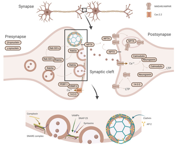
FIGURE 3.

Schematic illustration of the synaptic proteins, their cellular location, and their synaptic processes. The synaptic proteins in the panel collectively encompass the presynaptic (beta‐synuclein and gamma‐synuclein) and postsynaptic terminals (neurogranin) as well as proteins involved in vesicle trafficking (complexin‐2, syntaxins, VAMP‐2, rab GDI alpha, and AP‐2). Further, proteins native to both the presynaptic and postsynaptic terminal (14‐3‐3 and NPTX) involved in, among others, postsynaptic receptor regulation (NPTX and 14‐3‐3). AMPAR, α‐amino‐3‐hydroxy‐5‐methyl‐4‐isoxazolepropionic acid receptor; AP‐2, activating protein 2; Cav2.2, N‐type voltage‐gated calcium channel; CRMP‐2, collapsin response mediator protein‐2; NPTX, neuronal pentraxins; NMDAR, N‐methyl‐D‐aspartate receptor; PEBP‐1, phosphatidylethanolamine‐binding protein 1; rab GDI alpha, rab GDP dissociation inhibitor alpha; Rab3a, ras‐related protein 3a; SNARE, soluble N‐ethylmaleimide‐sensitive factor attachment receptor; VAMP‐2, vesicle‐associated membrane protein 2. Created with BioRender.com
