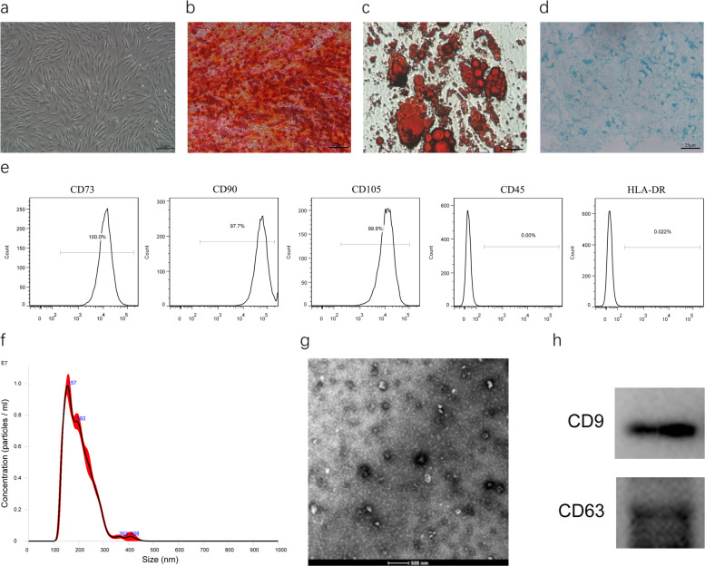
Fig. 1.
Isolation and characterization of hADSC and hADSC-Exo. a Spindle hADSC were observed through the microscope. Scale bar = 25 μm. b–d Adipogenic, osteogenic, and chondrogenic differentiation of hADSC. Cells differentiated into adipocyte, osteoblasts, and chondrogenic were detected using Oil Red O, Alizarin Red, and Alcian Blue, respectively. Scale bar = 25 μm. e Flow cytometry of hADSC surface markers CD29, CD73, CD105, SSEA-3, SSEA-4, and HLA-DR. f Nanoparticle analysis of ADSC-Exo. g TEM analysis of exosomes. Scale bars = 500 nm. h Immunoblotting for CD9 and CD63 in exosomes

