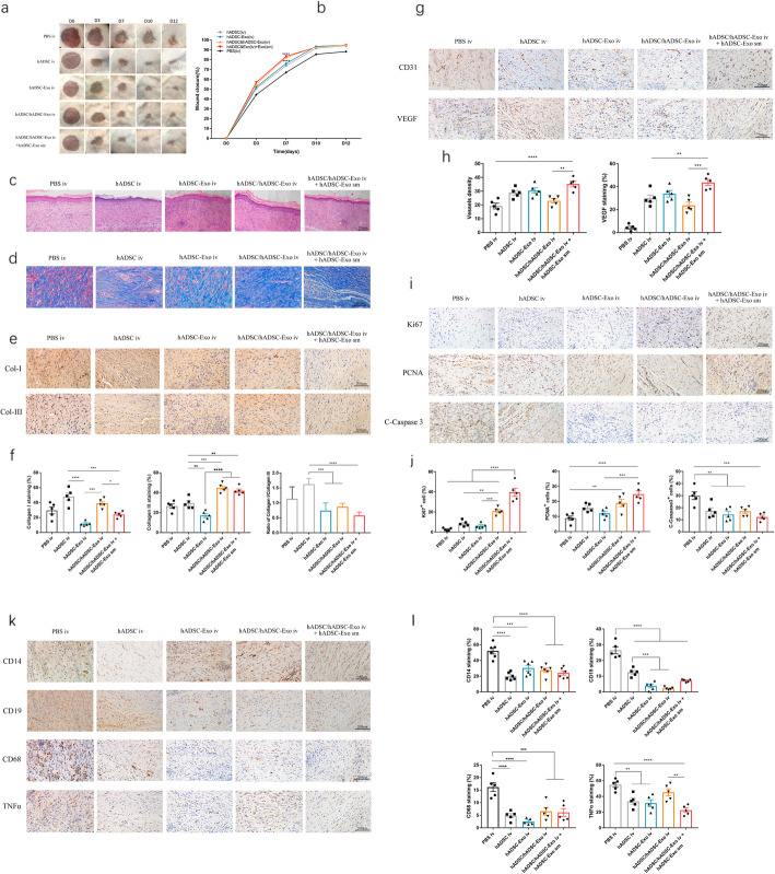
Fig. 5.
Therapeutic effects of hADSC-Exo in an in vivo mouse model. a Representative photographs of full-thickness excision wounds treated with hADSC iv and hADSC-Exo iv with or without hADSC-Exo sm. b Quantitative analysis of wound healing in each group. c Histological structure of wounded skin in different groups. Scale bar = 250 μm. d Masson staining of wounded skin sections in different groups at day 12 post-wounding. Scale bar = 200 μm. e The IHC of collagen-I and collagen-III in wounded skin sections on day 12 post-wounding. Scale bar = 200 μm. f Quantitative analysis of collagen I, collagen III, and the ratio of collagen I/collagen III. g Representative images of immunohistochemical results of CD31 and VEGF. Scale bar = 200 μm. h Quantitative analysis of the number of mature blood vessels. Quantitative analysis of the positive cells in the membrane tissues. i Representative images of immunohistochemical results of Ki67, PCNA, and C-Caspase3. Scale bar = 200 μm. j Quantitative analysis of the positive cells in the skin tissues. k Representative photographs of CD14, CD19, CD68, and TNF-alpha immunostaining. Scale bar = 200 μm. l Quantification of CD14+, CD19+, CD68+, and TNF-alpha+ IHC stained tissues. Results are presented as mean ± standard error of the mean; n = 5 for each group. *p < 0.05, **p < 0.01, ***p < 0.001, and ****p < 0.0001 vs vehicle control group

