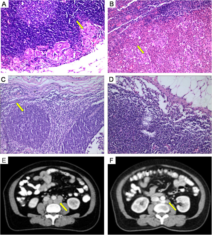
Fig. 2.
Computed tomography (CT) scans and postoperative hematoxylin and eosin (H&E) staining of responding lymph node. a. Lymph node metastasis of adenocarcinoma (yellow arrow) comfirmed by H&E staining. b. Lymph node metastasis of adenosquamous carcinoma (yellow arrow) comfirmed by H&E staining. c. Lymph node metastasis of squamous cell carcinoma (yellow arrow) comfirmed by H&E staining. d. Normal lymph nodes. e. Measurement of MAD (≥ 1.0 cm) of the para-aortic lymph node. The yellow arrows showed the location of lymph node metastasis. The H&E staining result was positive. f. Measurement of MAD (0.5–0.9 cm) of the para-aortic lymph node. The yellow arrows showed the location of lymph node metastasis. The H&E staining result was negative

