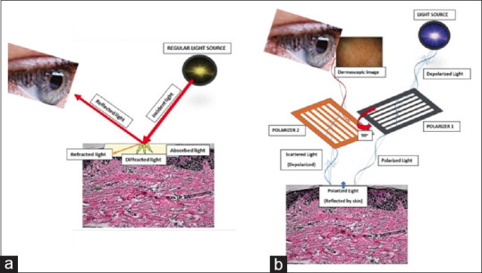
Figure 1.

(a) When light is incident on the skin (thick red arrow), it gets reflected back (thin red arrow), while the remaining gets refracted (oblique orange arrow), diffracted (yellow shooting arrows) or absorbed (crimson area). On looking directly at the skin by unassisted eyes, one sees the external image of the skin formed by the reflected light; (b) Working principle of a modern dermoscope. In the polarized mode, the light gets polarized by two cross-polarizers, cutting out the scattered light reflected from the skin, allowing image formation with visualization of substratal structures
