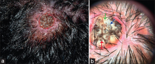
Figure 11.

Cutaneous Wound Myiasis: (a) Clinical image of wound myiasis showing well-circumscribed tender, boggy scalp ulcer with multiple larvae visible. Figure courtesy Gontijo and Bittencourt[52].(b) Dermoscopic image from the lesion showing numerous yellowish-white larvae with multiple brown dotted rings (green arrow), tracheal tube (blue arrow), and respiratory spiracle (red arrow). Erythema and perifollicular scaling on the scalp can be seen in the periphery. In the center, two bird's feet-like structures (white arrow) correspond to the breathing; Figure courtesy – Gontijo and Bittencourt[52]
