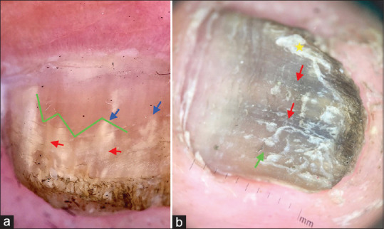
Figure 20.

Onychomycosis. (a) Jagged edge of the proximal margin of the onycholytic area (green lines), with sharp structure (spikes) directed to the proximal fold (blue arrows arrow), white- yellow longitudinal striae in the onycholytic nail plate (red arrows). (Dermlite DL3N 10X, polarized mode). (b) Fungal melanonychia: Dull matte black pigmented (red arrow) and yellow areas (green arrows) and pseudoleuconychia (yellow star). (Dermlite DL3N 10X, polarized mode)
