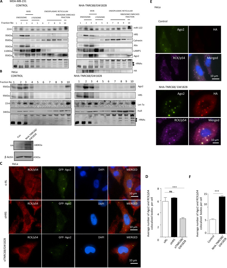
FIG 5.
GW182 proteins promote P-body targeting of miRNPs to restrict the miRNA export process. (A and B) Distribution of Ago2 and miRNA in MDA-MB-231 and HeLa cells expressing TNRC6B/GW182B. Increased miRNP is associated with endosome fraction positive for HRS marker protein in NHA-TNRC6B/GW182B-expressing MDA-MB-231 (A) and HeLa (B) cells. A 3 to 30% OptiPrep gradient analysis and distribution of Ago2 in cells expressing NHA-TNRC6B/GW182B are shown. Expression and distribution of NHA-TNRC6B/GW182B were determined by Western blotting using anti-HA antibody in panel A. Levels of let-7a or exogenously expressed miR-122 were determined by Northern blot analysis. Calnexin served as an ER marker. Alix was also an endosomal marker, while Lamp1 was used as a lysosomal marker. Distribution of stress response protein HuR was also determined in HeLa cells. (C) Inhibition of TNRC6B/GW182B by RNAi reduces Ago2-positive bodies in HeLa cells. Cells were transfected with GFP-Ago2 and siRNAs against TNRC6B/GW182B or HRS. GFP-Ago2 bodies were visualized in cells where they colocalized with RCK/p54. siRL was used as a control (means ± SEM; n = 20). (D) Quantification of Ago2 bodies showing colocalization with P-body marker RCK/p54 under different conditions (means ± SEM; n = 10 cells; P = 0.0005). (E) Larger numbers of P-bodies in NHA-TNRC6B/GW182B-expressing cells. HeLa cells were cotransfected with GFP-Ago2-encoding plasmid along with either the control or the NHA-TNRC6B/GW182B-encoding plasmids. GFP-positive Ago2 bodies were visualized and their colocalization with P-body marker RCK/p54 was documented. Increased size of Ago2 bodies showing colocalization with TNRC6B/GW182B was visualized in cells expressing NHA-TNRC6B/GW182B. (F) Relative quantification of colocalization of Ago2 and RCK/p54 from the experiments described for panel E was done and plotted (means ± SEM; n = 23 cells; P < 0.0001).

