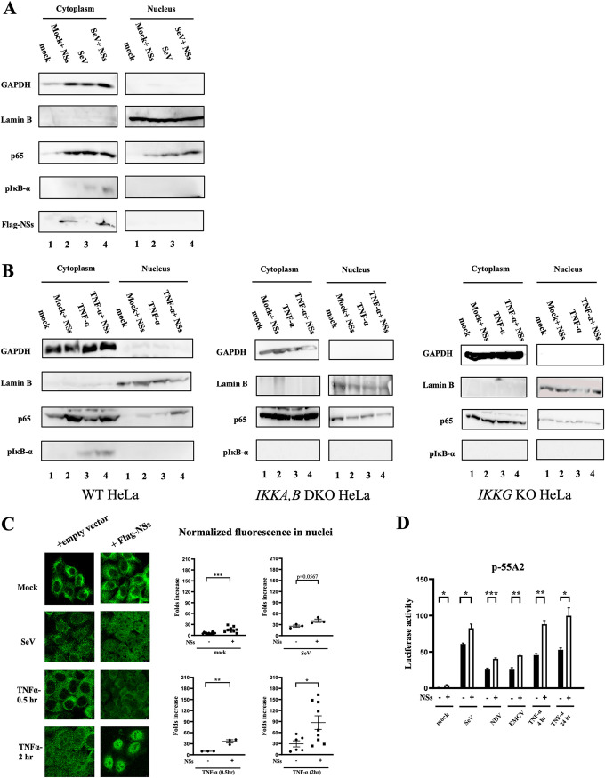
FIG 2.
NSs accelerated the activation of NF-κB. (A) HeLa cells (lanes 1 and 3) or those transfected with the expression vector for NSs-Flag (lanes 2 and 4) were mock treated (lanes 1 and 2) or infected with SeV for 9 h (lanes 3 and 4). The cells were subjected to subcellular fractionation. Cytoplasmic and nuclear extracts were examined by immunoblotting using the indicated antibodies. (A) A representative results of three independent experiments is shown. (B) Wild-type, IKKA,B DKO, and IKKG KO HeLa cells (lanes 1 and 3) or those transfected with the expression vector for NSs-Flag (lanes 2 and 4) were mock treated (lanes 1 and 2) or treated with recombinant TNF-α (20 ng/ml) for 30 min (lanes 3 and 4). The cells were subjected to subcellular fractionation. Cytoplasmic and nuclear extracts were examined by immunoblotting using the indicated antibodies. Comparison of nuclear p65 and cytoplasmic p-IκB-α is shown, with a representative result of two independent experiments for each cell line. (C) HeLa cells were transfected with the empty or expression vector for Flag-NSs. Twenty-four hours later, cells were mock treated or infected with SeV for 9 h or treated with TNF-α (20 ng/ml) for 0.5 or 2 h. Cells were fixed and stained for p65 for microscopy (left). Nuclear intensities were quantified (right). A representative result of two independent experiments is shown. (D) HEK293T cells were cotransfected with the NF-κB-dependent reporter gene p55A2 and pRL-TK for normalization with or without NSs-Flag. Twenty-four hours later, cells were mock treated or infected with different viruses (SeV, 9 h; NDV, 9 h; and EMCV, 12 h) or treated with TNF-α (20 ng/ml) for 4 or 24 h (n = 3). Luciferase activity was assessed using pRL-TK activity as a reference. Data are presented as means ± SEM. The Student t test was used for statistical analysis.

