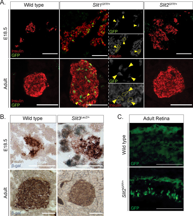
FIG 2.
Slit1, but not Slit2 or Slit3, is expressed in the mouse islet from embryonic stages to adulthood. (A) Immunofluorescence staining of β cells (insulin, red) and Slit1 and Slit2 (GFP, green) in E18.5 and adult heterozygous knock-in mice. Arrowheads indicate regions of overlapping GFP and insulin staining, representing Slit1-positive β cells. (B) β-Gal staining of Slit3 (LacZ, blue) in E18.5 and adult heterozygous knock-in mice. β-Gal staining (Slit3 expression) is apparent in nonendocrine tissue surrounding the islet in the embryo. Scale bar, 100 μm. (C) Immunofluorescence staining of Slit2 (GFP, green) in retinal sections from wild-type or Slit2GFP/+ heterozygous animals. Scale bars, 100 μm. n = 3 for all genotypes at each age analyzed.

