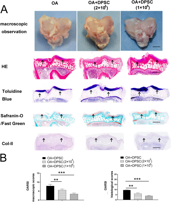
Fig. 6.
hDPSCs attenuated damage to the articular cartilage in vivo. The macroscopic characteristics of OA model group and the hDPSC treatment group are showed in a. In addition, the results of the HE, toluidine blue staining, safranin-O/Fast Green staining, and immunochemistry of Col-II showed that hDPSCs significantly improved the histological findings of osteochondral tissue in OA models. More intense staining in the cartilage layer was showed by using arrows, respectively (a). Moreover, the high-hDPSC group exhibited significantly lower pathological scores than the other 2 experimental groups (b). **P<0.01, ***P<0.001. Bars represent 3mm in microscopic data of a and 2mm in histopathological data in a, respectively. hDPSCs, human dental pulp stem cells; Col-II, collagen II; OA, osteoarthritis

