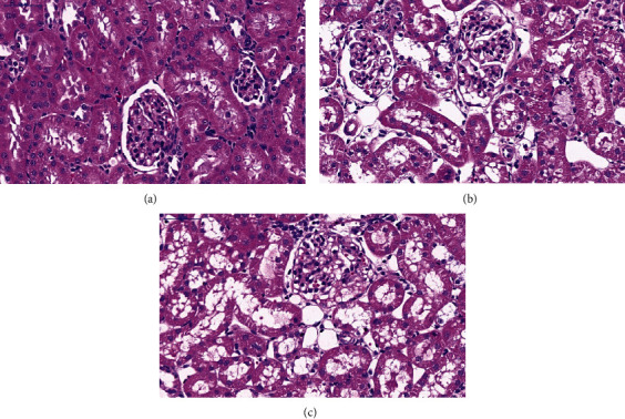
Figure 2.

Histological assessment of kidney injury in PBS- or LPS-treated rats by HE staining. (a) HE staining of the kidney tissue in the control (CT) group. (b) HE staining of the kidney tissue in the LPS2 group. (c) HE staining of the kidney tissue in the LPS6 group. HE staining demonstrated that renal tubular epithelial cells in the LPS2 (2B) and LPS6 (2C) groups were denatured and manifested vacuolar degeneration with detachment of the brush border, and the tubular lumen was enlarged compared with those in the control group (2A); necrotic shedding of epithelial cells and tubular formation were detected.
