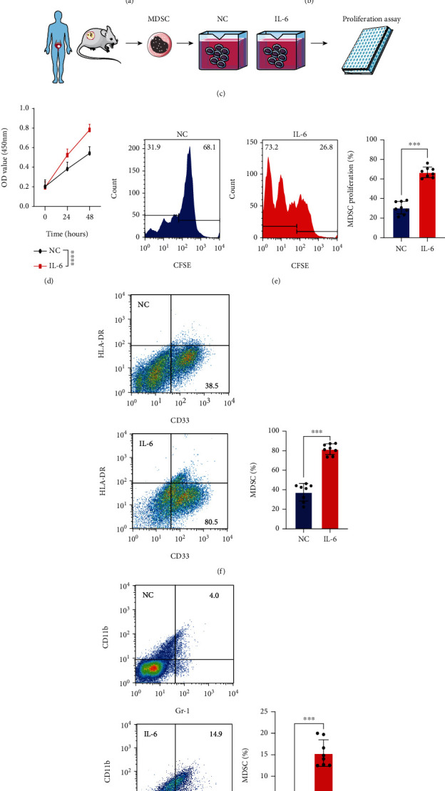
Figure 3.

IL-6 promoted MDSC proliferation in MIBC tissues. (a, b) Correlation between IL-6 (AOD and MFI) and MDSCs in MIBC tissues. (c) Experimental design of MDSC extraction and IL-6 treatment. (d) CCK-8 assays were used to test MDSCs with or without IL-6 treatment. (e) MDSC proliferation rates were evaluated according to fluorescence attenuation. (f, g) Flow cytometry images and statistical analysis of MDSCs from humans and mice with or without IL-6 treatment. Mean ± SD, ∗∗∗P < 0.005, and ∗∗∗∗P < 0.001. AOD: average optical density; MFI: median fluorescence intensity.
