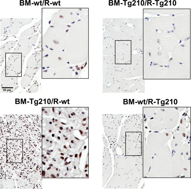
Fig. 4. Activation of TGF-β1-Smad3-P signaling pathway in ischemic BM-Tg210/R-wt gastrocnemius muscles.

Representative images of anti-phospho-Smad3 IHC of ischemic gastrocnemius muscle sections of BM-wt/R-wt, BM-Tg210-R-Tg210, BM-Tg210-R-wt, and BM-wt/R-210 chimeric mice. Positive nuclei are stained in brown/black (Smad3-P + hematoxylin), negative nuclei are stained in blue (hematoxylin alone). Magnification ×400, calibration bar 50 µm. Insets show magnifications of the indicated areas.
