
Fig. 5. α-SMA expression by BM-derived Tg-210 inflammatory cells.
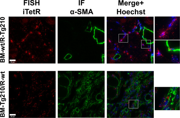
Representative images of double immunofluorescence staining for iTetR in situ hybridization (left, red), α-SMA (center, green) and merge (right) of ischemic gastrocnemius muscles sections of BM-wt/R-Tg210 mice and BM-Tg210/R-wt. Nuclei were stained by Hoechst (Blue). iTetR mRNA displays both nuclear and perinuclear staining. Representative images are presented as Maxprojections. Magnification 63 × 0.5, calibration bar 20 µm.