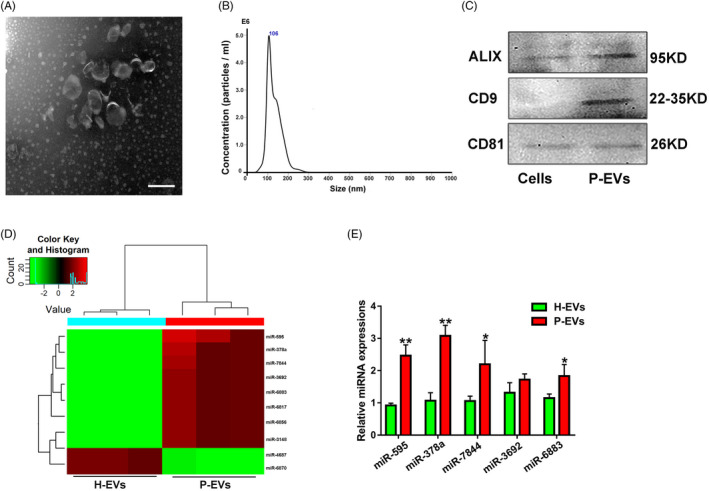
FIGURE 1.

Identification of an angiogenesis‐related miRNA (miR‐378a) differentially expressed in P‐EVs and H‐EVs. (A) Morphology of P‐EVs under transmission electron microscopy. (B) The size and concentration of P‐EVs were identified by nanoparticle analysis. (C) EV surface markers of ALIX, CD9 and CD81 detected by Western blot. (D) Heat map of differentially expressed miRNAs in P‐EVs and H‐EVs (fold change > 1.2, P <.05), wherein red colour represents a relative high expression level of the related miRNAs while green colour represents a relative low expression level of the related miRNAs. (E) Validation of the differentially expressed miRNAs screened from the Heat map in P‐EVs and H‐EVs by qRT‐PCR (n = 3); *P <.05 and **P <.01 vs. H‐EVs
