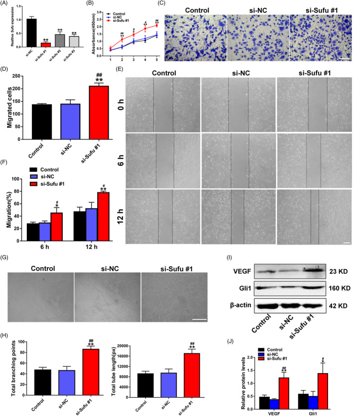
FIGURE 4.

Silencing of Sufu promotes EC proliferation, migration and angiogenesis via Hedgehog/Gli1 signalling activation. (A) The inhibitory efficiency of the siRNAs targeting Sufu was verified by qRT‐PCR. (B) The proliferation of ECs treated with PBS, si‐NC and si‐Sufu #1 was detected by CCK‐8 assay (n = 3). (C) The migration of ECs treated with PBS, si‐NC and si‐Sufu #1 was determined by transwell assay (scale bar, 100 μm). (D) Quantitative analysis of the migrated cells in C (n = 5). (E) The migration of ECs treated with PBS, si‐NC and si‐Sufu #1 was detected by scratch wound assay (scale bar, 200 μm). (F) Quantitative analysis of the migration rates in E (n = 5). (G) Representative images of the tube formation assay in ECs treated with PBS, si‐NC and si‐Sufu #1 (scale bar, 200 μm). (H) Quantitative analyses of the total branching points and total tube length in G (n = 3). (I) Detection of the protein level of VEGF in ECs by Western blot. (J) Quantitative analysis of the relative protein expression in I (n = 3). *P <.05, **P <.01 vs. the Control group; # P <.05, ## P <.01 vs. the si‐NC group
