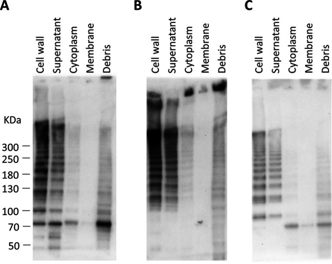
FIG 2.

Subcellular localization of pilus proteins in C. perfringens CP1. Total protein was separated by centrifugation and enzymatic treatment into five subcellular fractions and immunoblotting was performed using antiserum against CnaA (A), FimA (B), and FimB (C).
