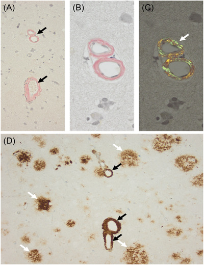
FIGURE 3.

Neuropathological study in individual II‐1 from this study (c.871A ≥ C, p.Thr291Pro). A, Several vessels with thickened vessels showing congophilia (black arrows; Congo Red staining). B, Higher magnification of A with congophilia of blood vessels (B) and after polarization of the same area showing typical apple‐green color (white arrow in C). D, Beta‐A4‐amyloid immunohistochemistry confirms amyloid deposition within the vessel walls (black arrows) and in addition several diffuse cortical plaques (white arrows), with the characteristic morphology of cotton‐wool plaques
