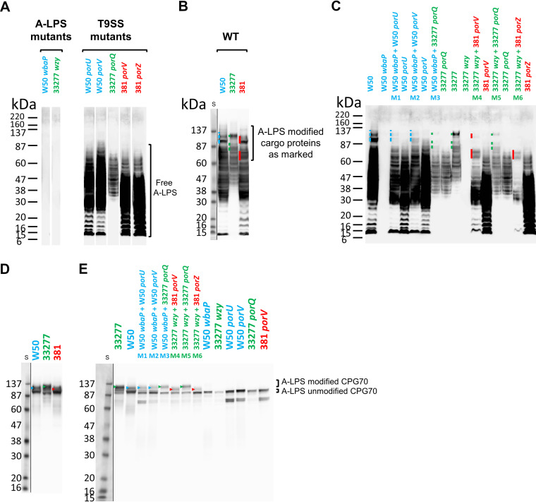
FIG 2.
Complementation of pigmentation on blood agar correlates with covalent linkage of protein to A-LPS. Whole-cell lysates of strains were grown for 6 days on plates as either single strains or mixes of an A-LPS mutant (W50 wbaP or 33277 wzy) with a T9SS mutant (W50 porU, W50 porV, 33277 porQ, 381 porV, or 381 porZ) as shown in Fig. 1B except for the WT strains (W50, 33277, and 381, shown in separate panels), which were similarly grown and processed. All strain names are colored according to strain background. (A, B, and C) Anti-A-LPS Western blot analyses. (D and E) Anti-CPG70 Western blot analyses. Profiles of high-MW A-LPS modified proteins are indicated by a colored line or arrowhead to the left of the band(s) and color coded according to the background strain (blue, W50; green, 33277; red, 381). S, protein size ladder. The resulting image of each of the Western blots showing the protein standard is produced by two exposures of the same blot perfectly overlapped and cropped to reveal the prestained protein ladder.

