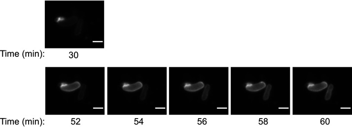
FIG 7.
Live capture of OMV-to-cell transfer of rhodamine B-R18 lipid dye. (Top) Close-up of two cells shown in Fig. 6E (30 min after mixing of wzy cells and rhodamine-labeled porU OMVs). (Bottom) Same cells showing a time-lapse series of rhodamine fluorescence every 2 min for 8 min. A single Z slice is presented. Bars, 2 μm.

