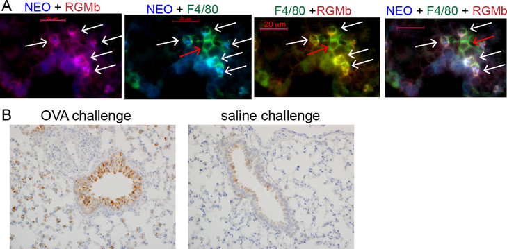
Figure 8.
NEO1 is expressed by RGMb+ interstitial macrophages and bronchiolar epithelium in lung tissue from OVA sensitized and challenged mice.
A. Left: Colocalization of NEO1 (blue) and RGMb (red) appears magenta (white arrows). 2nd panel: Colocalization of F4/80 (green) and NEO1 (blue) (white arrows). 3rd panel: Colocalization of F4/80 (green) and RGMb (red) appears yellow (white arrows). Right: Triple colocalization of NEO1, RGMb and F4/80 appears white (white arrows). Images representative of at least 4 animals. B. IHC staining of lung tissue for Neogenin. Bronchiolar epithelium from the saline challenged group shows patchy weak cytoplasmic staining. In the OVA challenged group, the bronchiolar epithelium shows diffuse strong cytoplasmic staining. NEO1-positive interstitial macrophages are also present in the OVA challenged lungs as compared to the saline treated mice.

