
FIGURE 2.
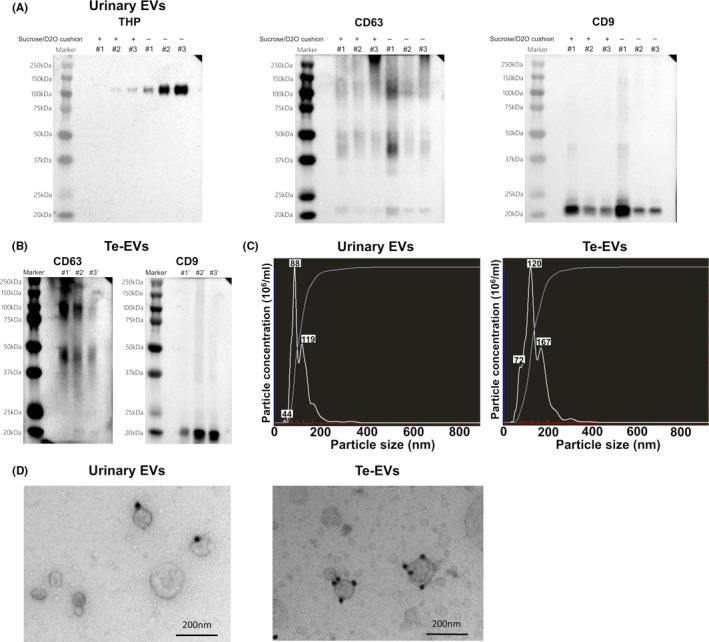
Verification of the quality of isolated urinary EVs and Te‐EVs. A, Western blot showing levels of EV‐specific proteins and THP in urinary EVs isolated using ultracentrifugation with or without a 30% sucrose/D2O cushion. B, Western blot showing levels of EV‐specific proteins in Te‐EVs. C, NTA revealing that almost all the extracted particles were <200 nm in size. D, Immunoelectron microscopy of urinary EVs and Te‐EVs immunolabeled with the anti‐CD9 antibody and secondary antibody conjugated to 20 nm of colloidal gold. BCa, bladder cancer; D2O, deuterium oxide; EVs, extracellular vesicles; NTA, NanoSight particle‐tracking analysis; Te‐EVs, tissue‐exudative extracellular vesicles; THP, Tamm‐Horsfall protein