FIGURE 3.

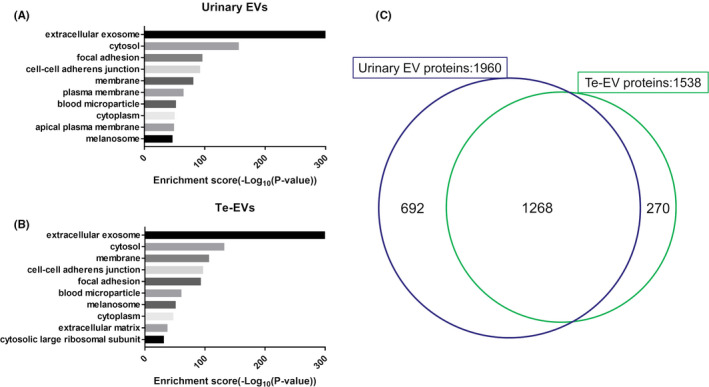
GO annotation (cellular component) of identified proteins using shotgun proteomic analysis in (A) urinary EVs and (B) Te‐EVs. C, Venn diagram showing the distribution of EV proteins identified using shotgun proteomic analysis. EVs, extracellular vesicles; GO, Gene Ontology; Te‐EVs, tissue‐exudative extracellular vesicles
