Figure 4.

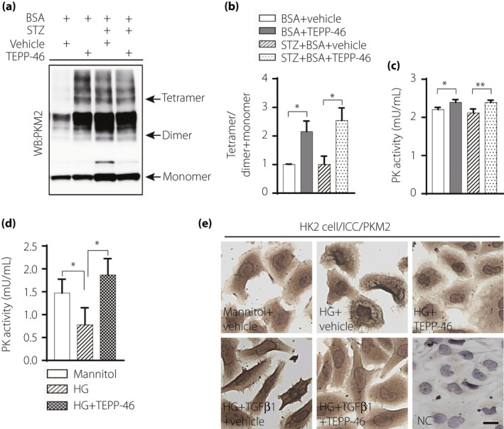
PKM2 activator TEPP‐46 increased PK activity, suppressed PKM2 nuclear location. (a) Representative blot image of cross‐linked mice kidney samples to show PKM2 monomer, dimer, and tetramer. (b) The ratio between tetramer and dimer + monomer of PKM2 in kidney samples, n = 5/group. (c) Analysis of pyruvate kinase (PK) activity in mice kidney samples, BSA, n = 5 mice; BSA + TEPP‐46, n = 6 mice; STZ + BSA, n = 8 mice; STZ + BSA+TEPP‐46, n = 7 mice. (d) HK2 cell treated by high‐glucose (30 mM) with TEPP‐46 for 48 h to analysis PK activity, n = 7 independent experiments. (e) Immunocytochemical analysis of PKM2 location in HK2 cell. (b, c) analyzed using unpaired two tail t‐test, (d) used one‐way ANOVA. The data are expressed as the mean ± SEM in the graph. P < 0.05 was recognized as significant. *P < 0.05, **P < 0.01. HG: high glucose.
