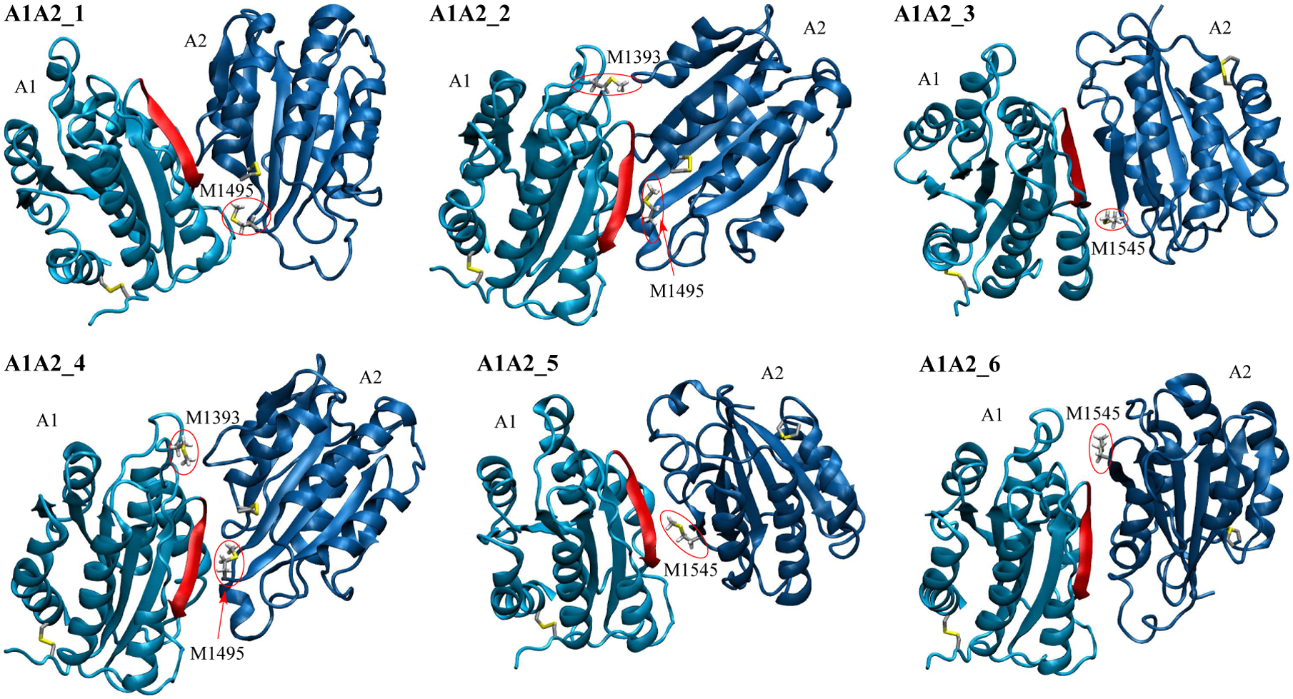
Figure 3: Models of the A1A2 complex.

Methionine residues located at the inter-domain interface (see “Materials and Methods”) and cysteine side chains forming disulfide bonds are shown in the stick and ball representation. Methionine residues at the interface are also labeled and highlighted by red circles. The backbone in the A1 domain colored in red highlights one of the major contact sites to GpIbα.56
