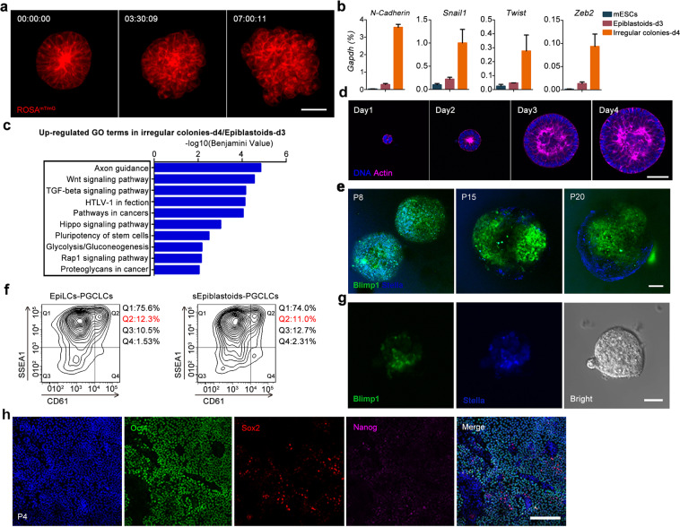
Fig. 2. Stabilizing and maintaining Epiblastoids by inhibiting Wnt/β-catenin activity.
a After culture for 3 days, ROSAmT/mG Epiblastoids (red) were recorded every 15 min for 24 h with Ultra VIEW-Vox system. Time-lapse images were processed with Velocity software 6.0. Scale bar, 40 μm. b qRT-PCR analysis of EMT-related markers N-cadherin, Snail1, Twist, and Zeb2 in mESCs, Epiblastoids at day 3 (Epiblastoids-d3), and irregular colonies at day 4 (irregular colonies-d4). The level of specific gene expression was normalized to Gapdh (100%). Data were represented as means ± SEM (n = 3). c GO analysis showed that the upregulated genes in day 4 irregular colonies were enriched in Wnt, TGF-beta and Hippo signaling pathways, compared with those in day 3 Epiblastoids. d mESCs differentiated into metastable Epiblastoids and were maintained in the medium with XAV939 (Epiblastoids medium) for 4 days. The different stages of cells were stained with Phalloidin (F-actin, magenta) and Hoechst 33342 (DNA, blue). Scale bar, 40 μm. e The Blimp1+ (green) and Stella+ (blue) cells displayed in the PGCLC aggregates of passage 8 (P8), 15 (P15) and 20 (P20) of Blimp1/Stella reporter sEpiblastoids at day 6 of differentiation. Scale bar, 100 μm. f FACS analysis of SSEA1 and CD61 (integrin-β3) double-positive cells for the aggregates of PGCLCs induced from P8-sEpiblastoids and EpiLCs at day 6 after differentiation. g PGCLCs (Blimp1+ and Stella+) displayed in the intact colonies of sEpiblastoids at day 6 after differentiation. Scale bars, 100 μm. h EpiLCs were induced from mESCs and propagated in sEpiblastoid medium in 2D culture condition. The immunofluorescent staining of Oct4, Sox2 and Nanog was performed for EpiLCs of passage 4 (P4). Scale bar, 200 μm.

