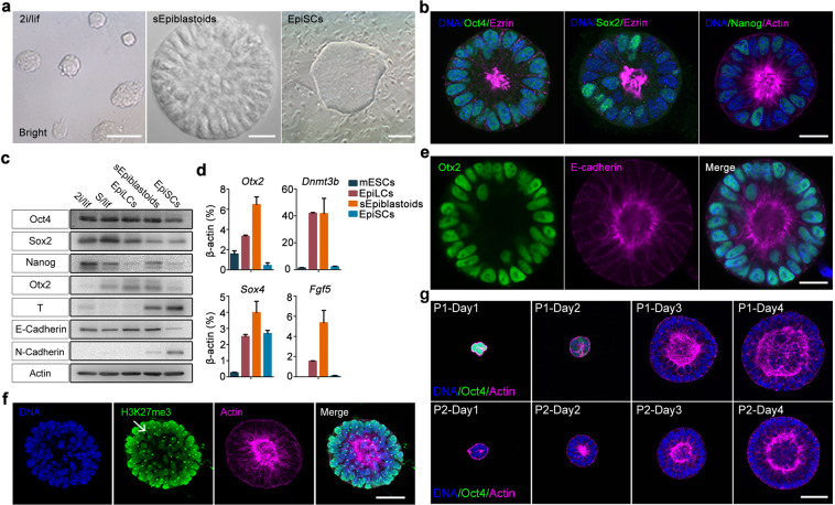
Fig. 3. Stabilized fPSCs possess features of formative pluripotent state.
a The morphology of typical naïve mESCs (left panel), sEpiblastoids (middle panel), and EpiSCs (right panel); scale bars, 50 μm (left panel), 20 μm (middle panel), and 300 μm (right panel). b The immunofluorescent staining of sEpiblastoids for Oct4, Sox2, or Nanog (green) and Ezrin or F-actin (magenta). DNA was stained with Hoechst 33342 (blue). Scale bar, 20 μm. c The lysates of mESCs (2i/lif or S/lif), 48 h EpiLCs, sEpiblastoids and EpiSCs were collected, and analyzed by western blot assay with specific antibodies for Oct4, Sox2, Nanog, Otx2, T, E-Cadherin and N-Cadherin. β-actin served as the loading control. d The expression of putative formative pluripotency markers (Otx2, Dnmt3b, Sox4 and Fgf5) was examined for naïve mESCs, EpiLCs, sEpiblastoids and EpiSCs by qRT-PCR with specific primers of these genes. The gene expression level was measured relative to β-actin (100%). Data were represented as means ± SD (n = 3). e sEpiblastoids were stained with specific antibodies for Otx2 (green), E-Cadherin (magenta), and Hoechst 33342 for DNA (blue). Scale bar, 20 μm. f Female fPSCs were stained with specific antibody for H3K27me3 (green) and Phalloidin for F-actin (magenta). DNA was stained with Hoechst 33342 (blue). Scale bar, 40 μm. g Oct4-ΔPE-GFP reporter mESCs were cultured in fPSC medium for 4 days (fPSCs passage 1, P1, top). P1 cells were propagated (P2, bottom) in the same medium. The cells of days 1–4 were stained with Phalloidin for F-actin (magenta) and Hoechst 33342 for DNA (blue). The enhancer utilization of Oct4 was indicated by GFP reporter (green). Scale bar, 40 μm.

