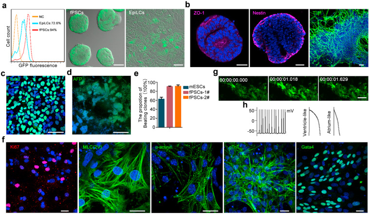
Fig. 6. fPSCs efficiently differentiate into all cell lineages of embryo in vitro.
a Sox1-GFP fPSCs and EpiLCs were derived from 46C mESCs and differentiated in neural induction conditions. After 2 days of differentiation, Sox1-GFP+ cells were analyzed by FACS. FACS analysis result was shown in left panel (NC, undifferentiated Sox1-GFP fPSCs). The photograph of Sox1-GFP+ cells were also shown. Scale bars, 100 μm (middle panel), 30 μm (right panel). b Neuronal lineage cells differentiated from fPSCs were stained with ZO-1 (tight junction protein 1, magenta) and Nestin (neural stem cell marker, magenta) antibodies at day 4, and Tuj1 (neuron marker, green) antibody at day 7 after differentiation. DNA was stained with Hoechst 33342 (blue). Scale bars, 50 μm. c fPSCs were induced into hepatocyte-like cells. At day 5 after differentiation, these cells were stained with Foxa2 (definitive endoderm marker, green). Nucleus was stained with Hoechst 33342. Scale bar, 50 μm. d The hepatocyte-like cells differentiated from fPSCs were stained with the antibody for AFP (liver cell marker, green) at day 12 after differentiation. DNA was stained with Hoechst 33342 (blue). Scale bar, 50 μm. e The proportion of beating cardiomyocyte-like cells induced from fPSCs and mESCs (EBs differentiation system). fPSCs-1#/2# represented two fPSCs lines (CMTI-1-fPSCs and 46C-fPSCs). All experiments were repeated at least three times. Error bars represented SEM. f The beating cardiomyocyte-like cells induced from fPSCs were stained with specific antibody for Ki-67 (proliferation marker), MLC(2V), α-actinin, cTnT and Gata4 (cardiomyocyte markers). Nucleus was revealed by Hoechst 33342 staining. Scale bars, 20 μm. g Time-lapse recording of Ca2+ releasing from the cardiomyocyte-like cells differentiated from fPSCs after 7–8 day differentiation. Scale bar, 40 μm. h Typical traces of simultaneous action potentials (APs) (left panel) and typical ventricle-like (middle panel) and atrium-like APs (right panel) observed in the cardiomyocyte-like cells differentiated from fPSCs after 7–8 days differentiation.

