
Fig. 1. Brain images of 4 patients with Hemorrhagic strokes presentation.
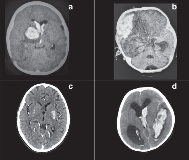
a basal ganglia hemorrhage, anterior horns of lateral ventricles; b right frontal hemorrhage, transtentorial herniation; c hypodensity in left basal ganglia, ischemic stroke, diffuse subcortical atrophy; d ventricular hemorrhage and hydrocephalus.