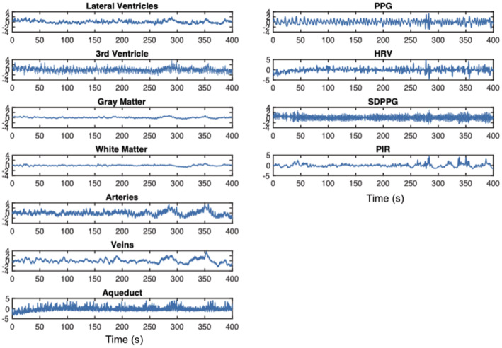
FIGURE 2.

Time series the fMRI in CSF‐related ROIs, contrasting signals from other ROIs as well as the PPG‐associated signals, from a representative subject. CSF‐related ROIs include the lateral ventricles (LV), the third ventricle (3rd V), and the cerebral aqueduct. All signals have been resampled (to the maximum frequency of the rs‐fMRI data). Column 1: It can be observed that the rs‐fMRI signals in the LV and 3rd V are synchronized with those in the arteries and veins, and to a lesser extent, those in the GM and WM. Column 2: It can also be observed that PIR best follows signals in these ROIs
