. 2021 Apr 9;24(2):196–205. doi: 10.4048/jbc.2021.24.e18

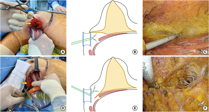
Figure 3. Illustrations of the flap dissections. (A) Images showing lifting of the lateral breast to approach the retromammary area (arrow, the lateral breast). (B) Schematic images showing the retromammary space dissection (yellow, glandular tissue; red, muscles in the axilla; blue, Glove Port; green, flexible endoscope). (C) Images showing the retromammary space dissection guided by a scope. (D) Images showing pulling of the lateral breast downward to approach the subcutaneous layer (arrow, the lateral breast). (E) Schematic images showing the subcutaneous flap dissection. (F) Images showing the subcutaneous flap dissection guided by a scope.

Figure 3