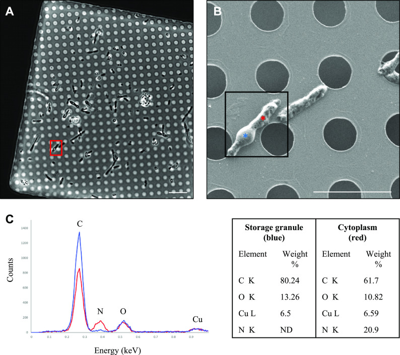
FIG 3.
Correlative LM and SEM of R. johrii for storage granule characterization with EDX. (A) An LM image of R. johrii shows the presence of storage granules (phase-bright objects) inside a cell (red square). (B) The same cell as in panel A imaged with SEM. Areas corresponding to the storage granule and cytoplasm are depicted by blue and red asterisks, respectively. (C) Elemental composition of the storage granule (blue) and cytoplasm (red) using EDX semiquantitative analysis. Major peaks are assigned and data are summarized in a table format. Scale bars, 10 μm (A) and 5 μm (B). ND, nondetected.

