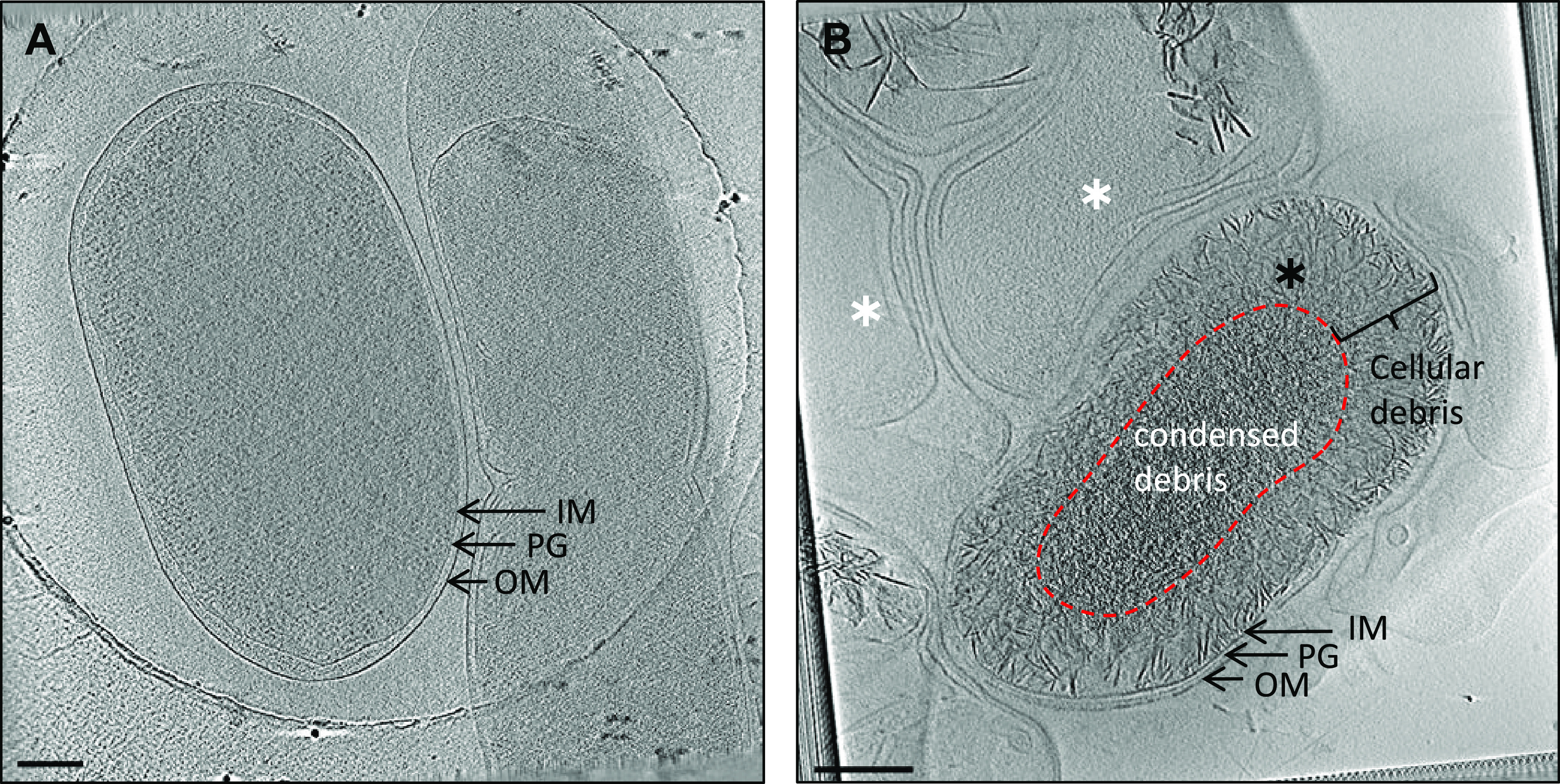
FIG 4.

Cryo-ET of S. marcescens. Tomographic slices through the following are shown: vegetative cells from a 2-day-old culture (A) and cells from a 65-day-old culture showing phase-bright objects (B). Panel B shows two cell types: cells with accumulated cellular debris (black asterisk) and ghost cells void of cellular material (white asterisks). Scale bar, 200 nm. IM, inner membrane; PG, peptidoglycan; OM, outer membrane.
