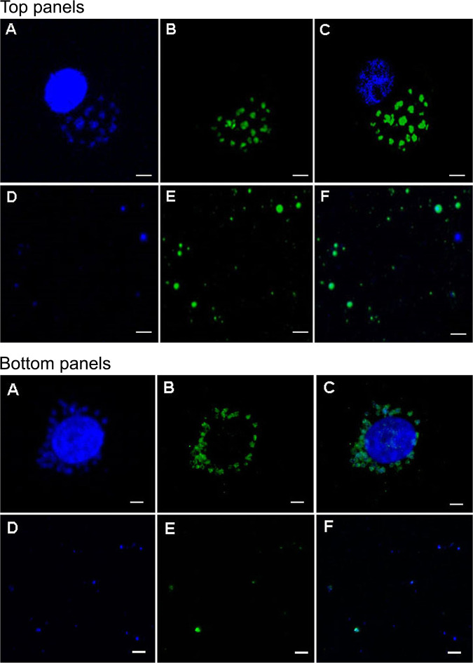
FIG 1.
(Top panels A to F) Representative confocal images showing phagosomes of the HL-60 cells infected by A. phagocytophilum stained with DAPI (A) and Rab 5 monoclonal antibody (B). A merged image is shown in panel C. Purified A. phagocytophilum phagosomes recovered from infected HL-60 cells were similarly stained with DAPI and Rab 5 antibody (panels D and E, respectively, with panel F representing the merged image). Scale bars in each panel are 5 μm. Bottom panels A to F present the data for E. chaffeensis cultured in DH82 cells. The image descriptions for bottom panels A to F are identical to descriptions for the top panels, including the scale bar size.

