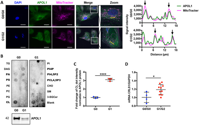
Figure 7 .

APOL1 is localized in mitochondria of urinary podocytes and binds with mitochondria-specific phospholipid cardiolipin. (A) Representative confocal images of differentiated podocytes stained with DAPI (blue), APOL1 (green) and MitoTracker (magenta). White color indicates overlapping pixels. Zoom: Regional magnification of the box area with the yellow line indicating the scanned segment (left panel, scale bar: 50 μm). Line scans of APOL1 and MitoTracker from left panel. The arrows represent colocalization (right panel). (B) APOL1 G0-6xHis and APOL1 G1-6xHis protein isolated from HeLa cells were overlaid in a lipid strip (upper panel). Purified APOL1-6xHis was confirmed by Western blot analysis (lower panel). (C) Scatter plots of the densitometric measurement of binding of Cardiolipin (CL) with APOL1 G0 and G1, normalized to 6xHis tagged APOL1 expression (n = 3). (D) Scatter plots of CRLS1 mRNA expression in G0/G0 and G1/G2 expressing podocytes (n = 4 & 10). The error bars represent mean ± SD of biologically independent experiments. Two-tailed Student’s t-test. *P < 0.05, ****P < 0.0001.
